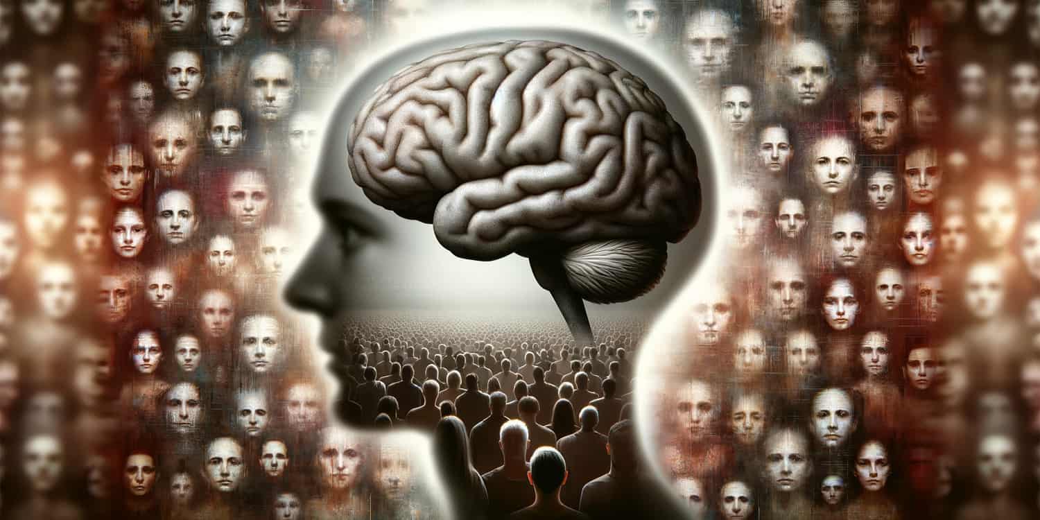
In a new study, Mayo Clinic researchers have uncovered new insights into prosopagnosia, commonly known as ‘face blindness,’ a condition where individuals struggle to recognize faces, including those of close family and friends. The research, published in Brain Communications, sheds light on the various neurological causes of this condition, its potential for improvement, and the specific brain regions implicated in its development.
What is Prosopagnosia?
Prosopagnosia is a neurological condition characterized by the inability to recognize faces. For individuals with prosopagnosia, the challenge is not in the basic visual processing of faces; they can see and often describe facial features. However, the brain’s ability to integrate these features into a recognizable whole face is impaired.
This can make it difficult to recognize close friends, family members, or even their own reflection. Prosopagnosia can be developmental, meaning a person is born with it, or acquired, resulting from brain injury or neurological diseases. The condition significantly impacts social interactions and emotional connections, as facial recognition is a key component of human communication and relationships.
Motivation Behind the Study
The researchers at the Mayo Clinic were motivated to conduct this study on prosopagnosia for several reasons. Firstly, there was a need for a clearer understanding of the different forms this condition can take. While the developmental type of prosopagnosia has been known, less was understood about the acquired form, particularly how it related to various neurological diseases and brain injuries.
“We were interested in knowing the extent to which different neurological diseases can be associated with prosopagnosia,” said study author Keith A. Josephs, a professor of neurology and neuroscience at the Mayo Clinic and co-author of “Common Pitfalls in Cognitive and Behavioral Neurology: A Case-Based Approach.”
Secondly, they aimed to delve into the specific brain regions associated with prosopagnosia. Previous studies had indicated involvement of certain areas like the right temporal or occipital lobes, but a more detailed exploration was necessary to confirm these findings and understand their implications.
Moreover, the researchers sought to investigate the potential variability in the presentation and progression of prosopagnosia. Particularly in acquired cases, there was curiosity about whether the condition could improve over time, depending on its underlying cause.
How the Study was Conducted
Initially, the Mayo Clinic’s Intake and Referral Center identified 487 patients with potential prosopagnosia from its campuses in Rochester, Minnesota; Scottsdale, Arizona; and Jacksonville, Florida, between January 2000 and January 2023. After rigorous screening, 336 patients were selected for the study based on specific inclusion criteria.
Patients were categorized into ‘definite’ or ‘probable’ prosopagnosia groups. Definite prosopagnosia was identified when patients had both self-reported difficulties and objective evidence from face recognition tests. Probable prosopagnosia was noted in cases where there was a subjective complaint but no formal testing or normal test results. The study also divided prosopagnosia into developmental and acquired types, the latter further split into degenerative (related to diseases that worsen over time) and non-degenerative categories.
Two types of face recognition tests were used: informal tests involving recognition of famous faces and formal tests requiring identification of a known face from a panel. The researchers also conducted thorough reviews of patient medical records to ascertain neurological diagnoses and reviewed neuroimaging data, including Magnetic Resonance Imaging (MRI) and Positron Emission Tomography (PET) scans.
Demographic and Clinical Characteristics
Among the 336 patients included in the study, a higher proportion were females (55%), with a median onset age for prosopagnosia of about 66 years. This indicates that while prosopagnosia can occur at any age, it predominantly emerges in the later stages of life, especially in the context of acquired prosopagnosia. Other visual agnosias were present in a quarter of these patients, highlighting the potential overlap with other neurological impairments.
Alternative Methods of Person Recognition
The researchers also observed that some patients with prosopagnosia had developed alternative strategies for person recognition, relying on senses other than sight. Notably, a number of individuals turned to auditory cues, like a person’s voice, or even olfactory cues, like their specific smell, to identify others.
“We were surprised at finding unusual behaviors employed by some of the patients with prosopagnosia, such as smelling a person, in order to recognize the individual,” Josephs said.
Developmental Prosopagnosia
A smaller subset, comprising 10 patients, was identified with developmental prosopagnosia. Intriguingly, this group had a notable male dominance (80%), and all were believed to be born with the condition. The study found genetic underpinnings in some of these cases, such as a mutation in the FOXG1 gene, indicating a potential hereditary aspect of developmental prosopagnosia. The discovery of these genetic links opens new avenues for understanding and perhaps diagnosing the condition early in life.
Acquired Prosopagnosia
The majority of the study’s participants had acquired prosopagnosia, with 235 cases attributed to neurodegenerative diseases and 91 to non-degenerative causes. This distinction is crucial as it underscores the different pathways through which prosopagnosia can develop, either as part of the progression of a degenerative neurological condition or due to an isolated incident like a brain injury or stroke.
Neurodegenerative vs. Non-Degenerative Causes
Among the neurodegenerative causes, conditions like posterior cortical atrophy, Primary Prosopagnosia Syndrome, and Alzheimer’s disease dementia were predominant. This finding is significant as it links prosopagnosia with well-known neurodegenerative diseases, suggesting that face-blindness can be a symptom of these broader disorders. On the other hand, in non-degenerative cases, the most common causes were strokes, epilepsy-related diseases, and brain tumors.
Neuroimaging Insights
In degenerative cases, the researchers found substantial involvement of bilateral temporal and occipital lobes, indicating these brain regions’ critical role in facial recognition processing. Conversely, in non-degenerative cases, lesions were predominantly observed in the right temporal or occipital lobes. This asymmetry is a crucial finding, suggesting a lateralized brain function in facial recognition in non-degenerative prosopagnosia cases.
Transient and Improving Prosopagnosia
A particularly intriguing aspect of the study was the observation that, in certain non-degenerative cases, prosopagnosia showed signs of improvement or was transient. This was especially noted in conditions like migraines and seizures. This discovery offers a glimmer of hope, suggesting that prosopagnosia may not always be a permanent condition and might improve depending on the underlying cause.
Together, these findings emphasize the complexity and diversity of prosopagnosia, highlighting various underlying causes and different patterns of brain involvement. They also offer hope that, in some instances, prosopagnosia may not be a permanent condition.
The study indicates “that there are many different causes of prosopagnosia both in childhood and adulthood and that prosopagnosia in adulthood is not always permanent depending on the cause,” Josephs told PsyPost.
Limitations and Future Directions
While study provides important insights, it’s important to note its limitations. Its retrospective design means it relied on existing medical records, which may not have uniformly captured all relevant data. Not all patients underwent standardized objective testing for prosopagnosia, and the testing methods evolved over the study’s 23-year span. “Not everyone in the study was formally tested with the identical test for loss of facial recognition,” Josephs explained.
Future research should focus on prospective studies with standardized testing protocols to assess prosopagnosia more consistently. Investigating the effectiveness of different treatment strategies and further exploring the genetic underpinnings of developmental prosopagnosia could also be beneficial.
“We plan to conduct future research to try to better understand the different types of prosopagnosia (associative and apperceptive),” Josephs said.
The study, “Prosopagnosia: face blindness and its association with neurological disorders“, was authored by Kennedy A Josephs and Keith A. Josephs, Jr.


